Abstract
Case Report
Cranio-Facial Fibrous Dysplasia: A case report of a conservative treatment in a monostotic form associated with an orthodontic management and a bone graft of the non-lytic bone area for dental implant rehabilitation
Seban A*, Blein E, Perez S and Seban B
Published: 21 November, 2019 | Volume 3 - Issue 1 | Pages: 018-022
Fibrous dysplasia is an osteolytic lesion in which bone is replaced by an instable fibrous osseous tissue. The aim of this case report is to highlight dental rehabilitation (bone grafts to allow dental implant) on patients suffering of this condition.
A 39-year-old female with a hard-traumatic event in childhood desired a dental implant rehabilitation on her teeth 19 and 30 after an orthodontic alignment. A Cone-Beam Computed Tomography (CBCT) was performed showing a massive radiopaque lesion of the anterior mandible. The bone grafts and dental implants were successfully managed. A non-invasive treatment with regular follow up was chosen for this case. No evolution was noticed twenty-four month later at the follow up CBCT.
Read Full Article HTML DOI: 10.29328/journal.jcad.1001011 Cite this Article Read Full Article PDF
Keywords:
Fibrous dysplasia; Monostotic cranio-facial dysplasia; Bone defect; Bone graft; Dental implant; Oral surgery
References
- Pack SE, Al Share AA, Quereshy FA, Baur DA. Osteosarcoma of the Mandible Arising in Fibrous Dysplasia-A Case Report. J Oral Maxillofac Surg. 2016; 74: 2229.e1-2229.e4. PubMed: https://www.ncbi.nlm.nih.gov/pubmed/27425882
- Qu N, Yao W, Cui X, Zhang H. Malignant transformation in monostotic fibrous dysplasia: clinical features, imaging features, outcomes in 10 patients, and review. Medicine (Baltimore). 2015; 94: e369. PubMed: https://www.ncbi.nlm.nih.gov/pubmed/25621678
- Leet AI, Collins MT. Current approach to fibrous dysplasia of bone and McCune-Albright syndrome. J Child Orthop. 2007; 1: 3-17. PubMed: https://www.ncbi.nlm.nih.gov/pubmed/19308500
- DiCaprio MR, Enneking WF. Fibrous dysplasia. Pathophysiology, evaluation, and treatment. J Bone Joint Surg Am. 2005; 87: 1848-1864. PubMed: https://www.ncbi.nlm.nih.gov/pubmed/16085630
- Hsissen MA, Kadiri F, Zamiati S, Jabri L, Rifki S, et al. A case of facial fibrous dysplasia in brothers. Rev Stomatol Chir Maxillofac. 1997; 98: 96-99. PubMed: https://www.ncbi.nlm.nih.gov/pubmed/9324737
- Hunter AG, Jarvis J. Osteofibrous dysplasia: two affected male sibs and an unrelated girl with bilateral involvement. Am J Med Genet. 2002; 112: 79-85. PubMed: https://www.ncbi.nlm.nih.gov/pubmed/12239726
- Parekh SG, Donthineni-Rao R, Ricchetti E, Lackman RD. Fibrous dysplasia. J Am Acad Orthop Surg. 2004; 12: 305-313. PubMed: https://www.ncbi.nlm.nih.gov/pubmed/15469225
- Sargin H, Gozu H, Bircan R, Sargin M, Avsar M, et al. A case of McCune-Albright syndrome associated with Gs alpha mutation in the bone tissue. Endocr J. 2006; 53: 35-44. PubMed: https://www.ncbi.nlm.nih.gov/pubmed/16543670
- Pierre Gangloff, Anne Polo, Hevré Moizan, Nicolas Froment, Eric Gerard. Maxillary tumefaction revealing a fibrous dysplasia. A case report. Med Buccale Chir Buccale. 2004; 10: 77-81.
- Long A, Loughlin T, Towers RP, McKenna TJ. Polyostotic fibrous dysplasia with contrasting responses to calcitonin and mithramycin: aetiological and therapeutic implications. Ir J Med Sci. 1988; 157: 229-234. PubMed: https://www.ncbi.nlm.nih.gov/pubmed/2971632
- Edgerton MT, Persing JA, Jane JA. The surgical treatment of fibrous dysplasia. With emphasis on recent contributions from cranio-maxillo-facial surgery. Ann Surg. 1985; 202: 459-479. PubMed: https://www.ncbi.nlm.nih.gov/pubmed/3901941
- Singh V, Gupta K, Salunke P. Monostotic craniofacial fibrous dysplasia: report of two cases with interesting histology. Autops Case Rep. 2019; 9. PubMed: https://www.ncbi.nlm.nih.gov/pubmed/31321219
- Kaynak BA. Conservative treatment of Fibrous Dysplasia. Pak J Med Sci. 2019; 35: 873-876. PubMed: https://www.ncbi.nlm.nih.gov/pubmed/31258611
- Majoor BCJ, Traunmueller E, Maurer-Ertl W, Appelman-Dijkstra NM, Fink A, et al. Pain in fibrous dysplasia: relationship with anatomical and clinical features. Acta Orthop. 2019; 90: 401-405. PubMed: https://www.ncbi.nlm.nih.gov/pubmed/31035847
- Kochanowski NE, Badry MS, Abdelkarim AZ, Lozanoff S, Syed AZ. Radiographic Diagnosis of Fibrous Dysplasia in Maxilla. Cureus. 2018; 10: e3127. PubMed: https://www.ncbi.nlm.nih.gov/pubmed/30345186
- Chennoju SK, Pachigolla R, Govada VM, Alapati S, Balla S. Idiosyncratic Presentation of Cemento-Osseous Dysplasia - An in Depth Analysis Using Cone Beam Computed Tomography. J Clin Diagn Res. 2016; 10: ZD08-10. PubMed: https://www.ncbi.nlm.nih.gov/pubmed/27437374
- Norris MA, Kaplan PA, Pathria M, Greenway G. Fibrous dysplasia: magnetic resonance imaging appearance at 1.5 tesla. Clin Imaging. 1990; 14: 211-215. PubMed: https://www.ncbi.nlm.nih.gov/pubmed/2224623
- Wilbur AC, Dobben GD, Linder B. Paraorbital tumors and tumor-like conditions: role of CT and MRI. Radiol Clin North Am. 1987; 25: 631-646. PubMed: https://www.ncbi.nlm.nih.gov/pubmed/3107049
- Casselman JW, De Jonge I, Neyt L, De Clercq C, D'Hont G. MRI in craniofacial fibrous dysplasia. Neuroradiology. 1993; 35: 234-237. PubMed: https://www.ncbi.nlm.nih.gov/pubmed/8459931
- Shah ZK, Peh WC, Koh WL, Shek TW. Magnetic resonance imaging appearances of fibrous dysplasia. Br J Radiol. 2005; 78: 1104-1115. PubMed: https://www.ncbi.nlm.nih.gov/pubmed/16352586
- Franz D, Wechselberger J, Rasper M, Specht K, Kehl V, et al. Milk cloud appearance-a characteristic sign of fibrous dysplasia on contrast-enhanced MR imaging. Eur Radiol. 2019; 29: 3424-3430. PubMed: https://www.ncbi.nlm.nih.gov/pubmed/31062134
- Johannsen A. Chronic sclerosing osteomyelitis of the mandible. Radiographic differential diagnosis from fibrous dysplasia. Acta Radiol Diagn (Stockh). 1977; 18: 360-368. PubMed: https://www.ncbi.nlm.nih.gov/pubmed/899860
- Leeds N, Seaman WB. Fibrous dysplasia of the skull and its differential diagnosis. A clinical and roentgenographic study of 46 cases. Radiology. 1962; 78: 570-582. PubMed: https://www.ncbi.nlm.nih.gov/pubmed/14463598
- Riddle ND, Bui MM. Fibrous dysplasia. Arch Pathol Lab Med. 2013; 137: 134-138. PubMed: https://www.ncbi.nlm.nih.gov/pubmed/23276185
- Alves N, de Oliveira RJ, Takehana D, Deana NF. Recurrent Monostotic Fibrous Dysplasia in the Mandible. Case Rep Dent. 2016; 2016: 3920850. PubMed: https://www.ncbi.nlm.nih.gov/pubmed/27340572
- Prada EJ, Hassan KH, Brandi ML, Falchetti A. Polyostotic form of fibrous dysplasia in a 13 years old Colombian girl showing clinical and biochemical response to neridronate intravenous therapy. Clin Cases Miner Bone Metab. 2009; 6: 264-265. PubMed: https://www.ncbi.nlm.nih.gov/pubmed/22461257
- Park BY, Cheon YW, Kim YO, Pae NS, Lee WJ. Prognosis for craniofacial fibrous dysplasia after incomplete resection: age and serum alkaline phosphatase. Int J Oral Maxillofac Surg. 2010; 39: 221-226. PubMed: https://www.ncbi.nlm.nih.gov/pubmed/20083386
- Xiang Fang, Hongyuan Liu, Yun Lang, Yan Xiong, Hong Duan. Fibrous dysplasia of bone: Surgical management options and outcomes of 22 cases. Mol Clin Oncol. 2018; 9: 98-103. PubMed: https://www.ncbi.nlm.nih.gov/pubmed/29977545
- Kos M, Luczak K, Godzinski J, Klempous J. Treatment of monostotic fibrous dysplasia with pamidronate. J Craniomaxillofac Surg. 2004; 32: 10-15. PubMed: https://www.ncbi.nlm.nih.gov/pubmed/14729043
- Lee JS, FitzGibbon EJ, Chen YR, Kim HJ, Lustig LR, et al. Clinical guidelines for the management of craniofacial fibrous dysplasia. Orphanet J Rare Dis. 2012; 7: S2. PubMed: https://www.ncbi.nlm.nih.gov/pubmed/22640797
- Kose TE, Dincer Kose O, Erdem MA, Cankaya A, Ozcan Duman I. Monostotic fibrous dysplasia presenting in maxilla: a case report. J Istanb Univ Fac Dent. 2016; 50: 38-42. PubMed: https://www.ncbi.nlm.nih.gov/pubmed/28955564
- Nair PP, Bhargava D, Thomas S, Shreenivas K. Immature fibrous dysplasia: a mixed radio-opaque radiolucent lesion. BMJ Case Rep. 2013; 2013. PubMed: https://www.ncbi.nlm.nih.gov/pubmed/23291823
Figures:
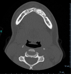
Figure 1

Figure 2

Figure 3
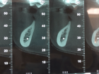
Figure 4

Figure 5

Figure 6
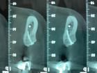
Figure 7
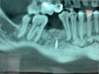
Figure 8

Figure 9
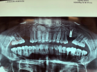
Figure 10
Similar Articles
-
Cranio-Facial Fibrous Dysplasia: A case report of a conservative treatment in a monostotic form associated with an orthodontic management and a bone graft of the non-lytic bone area for dental implant rehabilitationSeban A*,Blein E,Perez S,Seban B. Cranio-Facial Fibrous Dysplasia: A case report of a conservative treatment in a monostotic form associated with an orthodontic management and a bone graft of the non-lytic bone area for dental implant rehabilitation. . 2019 doi: 10.29328/journal.jcad.1001011; 3: 018-022
-
Bifid Mandibular Canals: A case report and mini reviewSourav Sarkar*,Saini Mondal. Bifid Mandibular Canals: A case report and mini review. . 2020 doi: 10.29328/journal.jcad.1001015; 4: 006-008
Recently Viewed
-
Agriculture High-Quality Development and NutritionZhongsheng Guo*. Agriculture High-Quality Development and Nutrition. Arch Food Nutr Sci. 2024: doi: 10.29328/journal.afns.1001060; 8: 038-040
-
A Low-cost High-throughput Targeted Sequencing for the Accurate Detection of Respiratory Tract PathogenChangyan Ju, Chengbosen Zhou, Zhezhi Deng, Jingwei Gao, Weizhao Jiang, Hanbing Zeng, Haiwei Huang, Yongxiang Duan, David X Deng*. A Low-cost High-throughput Targeted Sequencing for the Accurate Detection of Respiratory Tract Pathogen. Int J Clin Virol. 2024: doi: 10.29328/journal.ijcv.1001056; 8: 001-007
-
A Comparative Study of Metoprolol and Amlodipine on Mortality, Disability and Complication in Acute StrokeJayantee Kalita*,Dhiraj Kumar,Nagendra B Gutti,Sandeep K Gupta,Anadi Mishra,Vivek Singh. A Comparative Study of Metoprolol and Amlodipine on Mortality, Disability and Complication in Acute Stroke. J Neurosci Neurol Disord. 2025: doi: 10.29328/journal.jnnd.1001108; 9: 039-045
-
Development of qualitative GC MS method for simultaneous identification of PM-CCM a modified illicit drugs preparation and its modern-day application in drug-facilitated crimesBhagat Singh*,Satish R Nailkar,Chetansen A Bhadkambekar,Suneel Prajapati,Sukhminder Kaur. Development of qualitative GC MS method for simultaneous identification of PM-CCM a modified illicit drugs preparation and its modern-day application in drug-facilitated crimes. J Forensic Sci Res. 2023: doi: 10.29328/journal.jfsr.1001043; 7: 004-010
-
A Gateway to Metal Resistance: Bacterial Response to Heavy Metal Toxicity in the Biological EnvironmentLoai Aljerf*,Nuha AlMasri. A Gateway to Metal Resistance: Bacterial Response to Heavy Metal Toxicity in the Biological Environment. Ann Adv Chem. 2018: doi: 10.29328/journal.aac.1001012; 2: 032-044
Most Viewed
-
Evaluation of Biostimulants Based on Recovered Protein Hydrolysates from Animal By-products as Plant Growth EnhancersH Pérez-Aguilar*, M Lacruz-Asaro, F Arán-Ais. Evaluation of Biostimulants Based on Recovered Protein Hydrolysates from Animal By-products as Plant Growth Enhancers. J Plant Sci Phytopathol. 2023 doi: 10.29328/journal.jpsp.1001104; 7: 042-047
-
Sinonasal Myxoma Extending into the Orbit in a 4-Year Old: A Case PresentationJulian A Purrinos*, Ramzi Younis. Sinonasal Myxoma Extending into the Orbit in a 4-Year Old: A Case Presentation. Arch Case Rep. 2024 doi: 10.29328/journal.acr.1001099; 8: 075-077
-
Feasibility study of magnetic sensing for detecting single-neuron action potentialsDenis Tonini,Kai Wu,Renata Saha,Jian-Ping Wang*. Feasibility study of magnetic sensing for detecting single-neuron action potentials. Ann Biomed Sci Eng. 2022 doi: 10.29328/journal.abse.1001018; 6: 019-029
-
Pediatric Dysgerminoma: Unveiling a Rare Ovarian TumorFaten Limaiem*, Khalil Saffar, Ahmed Halouani. Pediatric Dysgerminoma: Unveiling a Rare Ovarian Tumor. Arch Case Rep. 2024 doi: 10.29328/journal.acr.1001087; 8: 010-013
-
Physical activity can change the physiological and psychological circumstances during COVID-19 pandemic: A narrative reviewKhashayar Maroufi*. Physical activity can change the physiological and psychological circumstances during COVID-19 pandemic: A narrative review. J Sports Med Ther. 2021 doi: 10.29328/journal.jsmt.1001051; 6: 001-007

HSPI: We're glad you're here. Please click "create a new Query" if you are a new visitor to our website and need further information from us.
If you are already a member of our network and need to keep track of any developments regarding a question you have already submitted, click "take me to my Query."


