Figure 1
Stability of facial soft tissue contour and bone wall at single maxillary tooth gap in early implant placement with contour augmentation: A case report
Cheng-Yi Wang and Jimmy LianPing Mau*
Published: 23 December, 2020 | Volume 4 - Issue 1 | Pages: 030-031
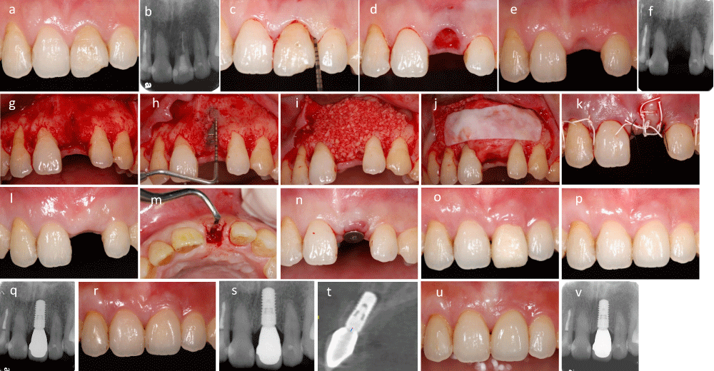
Figure 1:
1: (a,b): Root resorption of tooth 21 was diagnosed, (c): 3 mm bone sounding on buccal and mesiodistal aspect, (d): Less traumatic tooth extraction, (e,f): 2-week after tooth extraction, (g): Full thickness flap elevation, (h): 4.1 mm x10 mm Straumann bone level implant was placed in 3D position, (i-k): Freeze-dried bone allograft placed on the top of implant bony defect, two layers of collagen membrane was covered then primary closure was performed, (l-n): 3-month after implant placement, a U-shaped flap elevation for placement of healing abutment, (o): Provisional soft tissue modeling, (p,q): Final prosthesis delivery, clinical showed esthetic implant buccal hard and soft tissue contour, (r-t): 1-year post loading, clinical implant buccal hard and soft tissue contour remained stable, computed tomographic also showed even buccal bone wall, (u,v): 6-year follow up, clinical implant buccal hard and soft tissue contour still remained stable.
Read Full Article HTML DOI: 10.29328/journal.jcad.1001022 Cite this Article Read Full Article PDF
More Images
Similar Articles
-
Bilateral Parasymphyseal OsteomaAkanksha Gupta,Sangeeta Singh Malik,Swati Gupta,Ravi Prakash SM*. Bilateral Parasymphyseal Osteoma. . 2017 doi: 10.29328/journal.hjd.1001001; 1: 001-004
-
“Bulls Eye For Bulls Teeth”- Endodontic Management of Taurodontism Using CBCT as A Diagnostic Tool- 2 Rare Case ReportsSapna Sonkurla,Manoj Mahadeo Ramugade*,Shubha Hegde,Gopal Tawani. “Bulls Eye For Bulls Teeth”- Endodontic Management of Taurodontism Using CBCT as A Diagnostic Tool- 2 Rare Case Reports. . 2017 doi: 10.29328/journal.hjd.1001002; 1: 005-011
-
Diagnosis and Treatment of Anterior Cracked Tooth: A Case ReportWellington Luiz de Oliveira Da Rosa,Lucas Pradebon Brondani,Tiago Machado Da Silva,Evandro Piva, Fernandes Da Silva*. Diagnosis and Treatment of Anterior Cracked Tooth: A Case Report. . 2017 doi: 10.29328/journal.hjd.1001003 ; 1: 012-020
-
The Neuromuscular diseases in Pediatric Dental OfficeAmbarkova Vesna*. The Neuromuscular diseases in Pediatric Dental Office. . 2017 doi: 10.29328/journal.hjd.1001004; 1: 021-025
-
Fabrication of Lingual Retainer made easyMadhvi Bhardwaj, Shantanu Khattri,Rohit Kulshrestha*. Fabrication of Lingual Retainer made easy. . 2017 doi: 10.29328/journal.hjd.1001005; 1: 026-027
-
Staining susceptibility of recently developed resin composite materialsOlivier Duc,Emilie Betrisey, Enrico Di Bella, Ivo Krejci,Stefano Ardu*. Staining susceptibility of recently developed resin composite materials. . 2018 doi: 10.29328/journal.jcad.1001006; 2: 001-007
-
Preventing Peri-implantitis with a proper Cementation Protocol and with the consideration of alternatives to Cement-Retained Implant RestorationsTony Daher*,Robert G Mokbel, Vahik P Meserkhani. Preventing Peri-implantitis with a proper Cementation Protocol and with the consideration of alternatives to Cement-Retained Implant Restorations. . 2018 doi: 10.29328/journal.jcad.1001007; 2: 008-017
-
How Condylar modifications occursRohit Kulshrestha*. How Condylar modifications occurs. . 2018 doi: 10.29328/journal.jcad.1001008; 2: 018-019
-
Colour keys to emotion in management of children and intellectual distraction using coloured games in dental environment for childrenRajakumar S*,Kavitha Ramar, Revanth MP. Colour keys to emotion in management of children and intellectual distraction using coloured games in dental environment for children. . 2019 doi: 10.29328/journal.jcad.1001009; 3: 001-003
-
Cranio-Facial Fibrous Dysplasia: A case report of a conservative treatment in a monostotic form associated with an orthodontic management and a bone graft of the non-lytic bone area for dental implant rehabilitationSeban A*,Blein E,Perez S,Seban B. Cranio-Facial Fibrous Dysplasia: A case report of a conservative treatment in a monostotic form associated with an orthodontic management and a bone graft of the non-lytic bone area for dental implant rehabilitation. . 2019 doi: 10.29328/journal.jcad.1001011; 3: 018-022
Recently Viewed
-
Success, Survival and Prognostic Factors in Implant Prosthesis: Experimental StudyEpifania Ettore*, Pietrantonio Maria, Christian Nunziata, Ausiello Pietro. Success, Survival and Prognostic Factors in Implant Prosthesis: Experimental Study. J Oral Health Craniofac Sci. 2023: doi: 10.29328/journal.johcs.1001045; 8: 024-028
-
Agriculture High-Quality Development and NutritionZhongsheng Guo*. Agriculture High-Quality Development and Nutrition. Arch Food Nutr Sci. 2024: doi: 10.29328/journal.afns.1001060; 8: 038-040
-
A Low-cost High-throughput Targeted Sequencing for the Accurate Detection of Respiratory Tract PathogenChangyan Ju, Chengbosen Zhou, Zhezhi Deng, Jingwei Gao, Weizhao Jiang, Hanbing Zeng, Haiwei Huang, Yongxiang Duan, David X Deng*. A Low-cost High-throughput Targeted Sequencing for the Accurate Detection of Respiratory Tract Pathogen. Int J Clin Virol. 2024: doi: 10.29328/journal.ijcv.1001056; 8: 001-007
-
A Comparative Study of Metoprolol and Amlodipine on Mortality, Disability and Complication in Acute StrokeJayantee Kalita*,Dhiraj Kumar,Nagendra B Gutti,Sandeep K Gupta,Anadi Mishra,Vivek Singh. A Comparative Study of Metoprolol and Amlodipine on Mortality, Disability and Complication in Acute Stroke. J Neurosci Neurol Disord. 2025: doi: 10.29328/journal.jnnd.1001108; 9: 039-045
-
Development of qualitative GC MS method for simultaneous identification of PM-CCM a modified illicit drugs preparation and its modern-day application in drug-facilitated crimesBhagat Singh*,Satish R Nailkar,Chetansen A Bhadkambekar,Suneel Prajapati,Sukhminder Kaur. Development of qualitative GC MS method for simultaneous identification of PM-CCM a modified illicit drugs preparation and its modern-day application in drug-facilitated crimes. J Forensic Sci Res. 2023: doi: 10.29328/journal.jfsr.1001043; 7: 004-010
Most Viewed
-
Evaluation of Biostimulants Based on Recovered Protein Hydrolysates from Animal By-products as Plant Growth EnhancersH Pérez-Aguilar*, M Lacruz-Asaro, F Arán-Ais. Evaluation of Biostimulants Based on Recovered Protein Hydrolysates from Animal By-products as Plant Growth Enhancers. J Plant Sci Phytopathol. 2023 doi: 10.29328/journal.jpsp.1001104; 7: 042-047
-
Sinonasal Myxoma Extending into the Orbit in a 4-Year Old: A Case PresentationJulian A Purrinos*, Ramzi Younis. Sinonasal Myxoma Extending into the Orbit in a 4-Year Old: A Case Presentation. Arch Case Rep. 2024 doi: 10.29328/journal.acr.1001099; 8: 075-077
-
Feasibility study of magnetic sensing for detecting single-neuron action potentialsDenis Tonini,Kai Wu,Renata Saha,Jian-Ping Wang*. Feasibility study of magnetic sensing for detecting single-neuron action potentials. Ann Biomed Sci Eng. 2022 doi: 10.29328/journal.abse.1001018; 6: 019-029
-
Pediatric Dysgerminoma: Unveiling a Rare Ovarian TumorFaten Limaiem*, Khalil Saffar, Ahmed Halouani. Pediatric Dysgerminoma: Unveiling a Rare Ovarian Tumor. Arch Case Rep. 2024 doi: 10.29328/journal.acr.1001087; 8: 010-013
-
Physical activity can change the physiological and psychological circumstances during COVID-19 pandemic: A narrative reviewKhashayar Maroufi*. Physical activity can change the physiological and psychological circumstances during COVID-19 pandemic: A narrative review. J Sports Med Ther. 2021 doi: 10.29328/journal.jsmt.1001051; 6: 001-007

HSPI: We're glad you're here. Please click "create a new Query" if you are a new visitor to our website and need further information from us.
If you are already a member of our network and need to keep track of any developments regarding a question you have already submitted, click "take me to my Query."


